Internal carotid artery
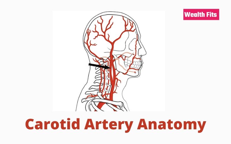
The internal carotid artery, which is distributed to the brain and organs of the visual apparatus. It originates from the common carotid artery at the height of the upper edge of the thyroid cartilage of the larynx and is directed up and back. So, It’s going up to reach the inferior orifice of the carotid canal of the temporal bone. It traverses the channel, following its curvatures, and emerges from the upper orifice of the canal itself. Here you can know about carotid artery anatomy. It was thus penetrating the cranial cavity. It then goes inside the cavernous sinus, within which it runs with a Sway, at the height of the anterior clinoid process.
The artery becomes vertical and perforates the dura mater. Here it provides voluminous collateral, the ophthalmic artery. Then ends, below the anterior perforated substance of the brain. It is dividing into the anterior cerebral, middle cerebral, anterior choroid, and posterior communicating arteries, which are its terminal branches.
In the neck, the internal carotid artery is located first behind and laterally to the external carotid artery whose relationships it shares. The stylohyoid muscle and the posterior belly of the digastric muscle. Having thus acquired a profound situation.

Internal Carotid
The internal carotid artery relates to: forward, with the pharyngeal extension of the parotid gland. Behind the prevertebral muscles and the cervical chain of the orthosympathetic that are behind the deep cervical fascia. Medially, with the lateral wall of the pharynx, laterally, with the styloglossus muscle and the stilopharyngeal muscle and with the glossopharyngeal nerve. The internal carotid artery is accompanied, in its course,
In the carotid canal, the internal carotid artery is surrounded by the carotid plexus of the orthosympathetic. With the intermezzo of the bony walls of the channel. Enters into a relationship with the cavity of the tympanum laterally, with the auditory tuba forward, with the cochlea in behind. The semilunar ganglion of the trigeminal rests on the upper wall of the horizontal section of the carotid canal.
He was released from the carotid canal. The artery runs over the fibrocartilage, which obliterates the lacerated hole and folds upwards, penetrating the cavernous sinus. Inside the breast, it is wrapped, on the surface, by a layer of endothelium and is surrounded by the nerve filaments of the Orth sympathetic that make up the cavernous plexus. In the same section, it is also related to the abducent nerve that runs outside and with the oculomotor. Trochlear and ophthalmic nerves that run in the lateral wall of the cavernous sinus. On exiting the cavernous sinus, the internal carotid crosses the optic nerve laterally, crosses the arachnoid, and reaches its termination.
The internal carotid artery does not provide collateral branches in the neck.
In the carotid canal it gives the following collateral branches:
- Carotidympanic branch, which crosses the posterior wall of the carotid canal and enters the hollow of the eardrum.
- Pterygoid branch, inconstant, which enters the pterygoid canal with the homonymous nerve. Cavernous units, for the semilunar ganglion and the walls of the cavernous sinus.
- Pituitary branches, for the pituitary and the ventral part of the hypothalamus. In the cranial cavity, it provides a single collateral branch, the ophthalmic artery.
Terminal branches of the internal carotid artery are the anterior choroidal artery, the posterior communicating artery. The middle cerebral artery and the anterior cerebral artery.
Common carotid artery
The common carotid arteries are the main arterial vessels for spraying the head and neck. The right common carotid artery detaches from the brachiocephalic trunk, behind the right sternoclavicular joint. The left common carotid artery originates directly from the arch of the aorta. Both arteries arise in the neck. Up to the height of the upper edge of the thyroid cartilage of the larynx, where they end dividing each into an external carotid artery. Face and upper part of the neck and an internal carotid artery
that irrigates the organs contained in the cranial cavity and the orbital cavities. Each common carotid artery has, at its upper end, near the bifurcation, an expansion. The carotid sinus, which, mostly extends to the initial tract of the internal carotid artery.
Carotid Sinus
In correspondence with the carotid sinus, the arterial wall presents a somewhat thickened adventitia which welcomes numerous nerve endings from a branch of the glossopharyngeal nerve. Due to its wealth of nerve endings, the carotid sinus acts as a baroreceptor, i.e., organelle sensitive to changes in blood pressure. In the bifurcation corner of the common carotid artery, there is a reddish-brown corpuscle, the gloom (or paraganglion) carotid, which has functions of chemoreceptor as warns of changes of the chemical composition of the blood.
In the thorax, the left common carotid artery is located at a certain distance from the sternal handlebar, from which the left brachiocephalic venous trunk that crosses it anteriorly separates it. Back, it has a relationship with the left subclavian artery, with the left edge of the trachea and, behind it, with the left edge of the esophagus, along which the left lower laryngeal nerve runs. Its medial margin is originally related to the brachiocephalic trunk; moving upwards, the two vessels diverge and thus delimit a space in which the trachea is included. The left vagus nerve runs along the lateral margin of the thoracic tract of the left common carotid artery and heads towards the aortic arch.
Carotid Arteries
In the neck, the common carotid arteries, right and left, have identical ratios. Each artery runs behind the sternocleidomastoid muscle together with the internal jugular vein, which is placed laterally and the vagus nerve that runs posteriorly to the two vessels. Together, these three formations wrapped in a connective sheath make up the vascular-nervous bundle of the neck.
However, about halfway down the neck, the common carotid artery is crossed by the upper belly of the hominoid muscle; below this, the artery is covered by the middle cervical fascia; above the muscle, it has a relationship with the descending branch of the cervical loop of the hypoglossal nerve and with the upper cardiac branches of the vagus nerve. Back the artery enters into a relationship, through the deep cervical fascia, with the cervical chain of the orthosympathetic, with the prevertebral muscles and with the transverse processes of the cervical vertebrae; the anterior tubercle of 6 to the cervical vertebra (or tubercle Chassaignac) is a landmark for the common carotid artery. At the medial side of the common carotid artery are the trachea, below, the larynx and the pharynx, above.
The common carotid artery does not give collateral branches. Its terminal branches are the external carotid artery and the internal carotid artery.
Online Yoga By Glo Is A Stress Buster
Carotid stenosis
Carotid stenosis is a disease that affects the carotid arterial system.
The term stenosis, in this case, indicates a reduction in the vascular caliber. As a result of which the blood flow downstream of the narrowing is decreased; this results in a state of suffering of the organs reached by it due to a lack of oxygen and nutrients transported by the blood. Carotid-stenosis Since the carotid artery irrigates the cerebral districts, the face, and the eyes, carotid stenosis causes the suffering of these anatomical areas and not only; in fact, the functionality of the limbs innervated by the affected brain areas is also impaired.
The main cause of carotid stenosis is atherosclerosis, a particular form of arteriosclerosis that affects large vessels.
Firstly, the main cause of carotid stenosis is atherosclerosis, a particular form of arteriosclerosis, which preferably affects large-caliber arterial vessels. So, at the level of the intimate tunic and of the innermost layers of the middle tunic of the arterial vessel, of a raised plaque with precise contours. This focus is called atheroma. Atheroma has a fibro lipidic consistency: the fibrous component is due to a proliferation of fibrous connective tissue (“scar” tissue); the lipid component comes. However, from the blood plasma and consists of cholesterol crystals, triglycerides, and fatty acids.
The onset of an atheroma is due to several factors, all equally important. The best known are:
- Hypertension
- Obesity
- Smoke
- Hypercholesterolemia
- Sedentary life
- Diabetes
- Aging
The atheroma, which develops at the level of the intimate tunic of the vessel, arises following an imbalance between the vessel wall and blood circulating in the lumen of the artery. That is, in the endothelium. The lesion creates an inflammatory situation and attracts cells of the plasma blood, such as red blood cells and white blood cells, whose intervention generates the first small plaque.
Carotid Artery Anatomy: Hypertension
For example, it creates a swirling flow within the arteries. This explains why atheromas develop electively where there are bifurcations of the carotid artery. Another example of instability in the relationship between the internal wall of the carotid artery and the blood concerns aging, an event that affects every individual. It reduces the elasticity and contractility of the arteries, thus changing their blood flow.
The picture is also enriched with the formation. So, at the level of atheroma, of a thrombus. So, the thrombus is a solid mass of blood cells. The consequence is natural. These actors combine to increase the thickening of the atheroma. At this point, the lumen of the arterial vessel of the carotid artery further narrows.
To worsen the situation even more. So, it is the possibility that the thrombus splits into smaller particles. In other words, these free particles, called emboli, can reach the brain. They are accelerating the processes of cerebral ischemia and stroke.
Other causes of carotid stenosis are:
- Aneurysms
- Fibromuscular dysplasia’s
- arteritis
- kinking
- Coiling
Carotid Artery Anatomy: Symptoms and signs
A clinical sign of carotid stenosis is the absence of pulsations in the affected vessel. In fact, the pulsation may also be present in conjunction with a narrowing of the carotid artery.
The main sign that characterizes carotid stenosis is the so-called transient ischemic attack, also known as TIA. The ischemic attack occurs at the cerebral, facial, and ocular levels, that is, the areas not sufficiently sprayed by the occluded carotid artery.
- Loss of control of the limbs: hemiplegia on the side opposite to that of the occluded carotid artery. Sprayed by the right carotid, controls the limbs of the left part of the body.
- Difficulty in speaking: language sometimes becomes incomprehensible.
- Vision problems: doubled or blurred vision. Possible blindness, which initially presents itself with a black or gray veil that drops before the eye. In this case, the affected eye is located on the same side of the occluded carotid artery.
- Lack of coordination in walking.
- Paresis of the face.
Carotid Artery Anatomy: Stenosis
Firstly, if stenosis involves major ischemic damage, which lasts up to 3 days, it’s reversible ischemic neurological deficits. The symptoms are similar to those of the TIA.
If, finally, the occlusion of the carotid artery is severe and almost. If not completely, complete. The symptom that derives from it is the ischemic stroke or stroke. In other words, the consequences are evident and no longer transitory. In most cases, this situation leads to death.
Carotid Artery Anatomy: Diagnosis
Firstly, the diagnosis of carotid stenosis can be based on the monitoring, through simple palpation, of the pulsations of the carotid artery. So, the absence of pulsation at the level of one of the two carotids could mean that an occlusion exists.
An important test is the so-called carotid sign. It consists of alternately compressing one of the two carotids and interrupting the blood flow that flows through the carotid vessel. Suppose the compressed carotid is the healthy one, after a time varying from 10 to 30 seconds. So, the patient shows signs of malaise, pallor, and loss of consciousness. The patient does not show symptoms. Since the opposite way, patent, compensates for the lesser flow, due to the stenosis, to the cerebral districts.
Instrumental diagnostic tests consist of:
- eco doppler
- Digital angiography
- Angioscanner
- Angio
What is DASH Diet and How the DASH Diet Can Help Your Cholesterol and Blood Pressure