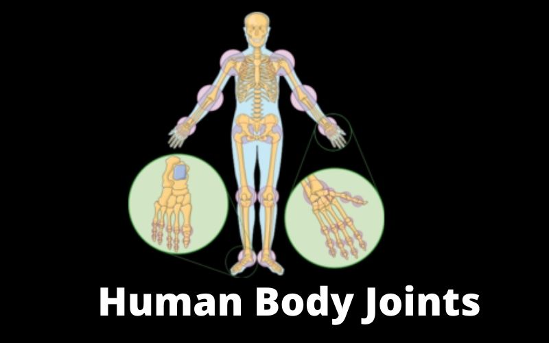
Hip (or coxo-femoral) joint
The hip joint (or coxo-femoral) is a typical enarthrosis (mobile joint – diarthrosis -, the articular surfaces of which consist of a spherical or hemispherical shape and a concavity which is, in turn, spherical) which joins the femur to the hip bone. The hip bone concurs with an almost hemispherical articular cavity. Here you can know about human body joints and the femur with the femoral head, which represents about 2/3 of a full sphere of 4 or 5 cm in diameter.
Similarly to what happens in the homologous shoulder joint (or shoulder joint), the joint surfaces are not perfectly corresponding. A glenoid rim, the lip of the acetabulum, widens the surface of the cavity and makes it suitable for containing the head of the femur. Unlike the glenoid lip of the scapulohumeral joint, which has no other function than that of enlarging the corresponding cavity, the acetabular lip has an important role in the union between the femur and the hip; it is, therefore, a means of containing the joint. The acetabular lip also bridges the notch of the acetabulum, converting it into a hole.
Glenoid cavity
Not all the glenoid cavity takes a direct part in the joint; in its center there is a quadrilateral depression, the fossa of the acetabulum, not covered with articular cartilage, but with a periosteum. From this fossa starts a ligament, with a rectangular section, the round ligament of the femur, which ends up on the fovea capitis of the femoral head and which, as a rule, does not exceed 35 mm in length.
The joint capsule is a fibrous sleeve, inserted proximal on the contour of the acetabulum and the acetabular lip and distally on the interchangeable line, forward, and a line placed on the border between the middle third and lateral third of the femoral neck, behind. In this way, the anterior face of the anatomical neck of the femur is intramuscular, while the posterior face is only in the 2/3 medial.
The longitudinal, memorabilia, ischiofemoral, and pubofemoral reinforcement ligaments are not dissociable from the capsule.
Iliofemoral ligament
The iliofemoral ligament is fan-shaped; it originates below the anterior inferior iliac spine, with two bundles that diverge like a fan. The oblique beam, directed to the anterior margin of the greater trochanter and the vertical beam. Towards the lower part of the intertrochanteric line.
- The pubofemoral ligament arises from the pubic tract of the edge of the acetabulum. The ischiofemoral ligament is triangular, and from the ischial side of the cotyloid eyelash, it leads out to the trochanteric fossa.
- The orbicular area, covered by the previous ligaments. Detaches from the edge of the acetabulum and the acetabular lip. Deeply to the insertion of the iliofemoral ligament and, passing behind the neck of the femur that embraces the loop. Returns to fix itself at the point of origin.
- The round ligament of the femur extends from the fovea capitis. From which it descends, widening and remaining applied on the head of the femur, to then reach, with two roots—the edges of the notch of the acetabulum. Flat and laminar, the round ligament is not as tense as interosseous ligaments usually are.
Synovial
The synovial presents the characteristic disposition of diarthrosis. It covers the internal surface of the capsule. Having reached its insertions, it is reflected with a recurring route to coat the intracapsular portions of the bone heads up to the limits of the articular cartilages. It forms a complete sheath to the round ligament.
Knee joint
Anatomy
The knee is an articulation in which the femur, tibia, and patella come into play.
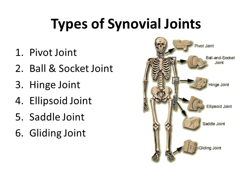
It is difficult to classify because of the joint relationship established between the femur. Patella can be defined as an arthrodia, while the femorotibial one is attributable. For some characters, to the condyloid joints. Others to the angular gingles. Furthermore, while the articular surfaces seem to allow extended freedom of movement, the ligamentous apparatus of the joint ends up limiting them to the only flexion-extension. At the level of the knee, there is the transmission of body weight to the leg: the joint, therefore, also has an important static task.
The femur participates in the joint with the anterior patellar surface. The two sides of the trochlea diverge posteriorly, and a deep intercondyloid notch follows the throat. However, the sides of the notch. The posterior extensions of the trochlea form two convex reliefs in the anteroposterior direction, the condyles. The tibia takes part in the joint with the upper end. They are opposing the two glenoid cavities on the internal and external tibial condyles to the femoral condyles. So, the glenoid cavities have shallow oval surfaces and are separated by a non-articular. The rough area that rises in the middle to form the intercondyloid eminence and widens backward and forwards in the respective intercondylar pits.
The patella participates in the joint with its posterior surface that corresponds to the femoral trochlea.
Human Body Joints: Menisci
The marked sagittal convexity of the two femoral condyles does not correspond to an equal concavity of the tibial surfaces. The harmony between the articular surfaces is therefore established by the interposition of two menisci. One medial and one lateral. These have the shape of half-rings, and their thickness is reduced, proceeding from the periphery to the center. Seen in section, they, therefore, have a triangular profile.
The lateral meniscus forms an almost complete circle. The medial one is interrupted on the internal side and therefore has the shape of C. With their ends (or horns), the menisci are fixed on the intercondylar portion of the tibia. The transverse knee ligament.
Joint capsule
Firstlty, the fibrous layer of the joint capsule constitutes a sleeve characterized by the brevity and solidity of the lateral and posterior parts and by the laxity of the anterior portion. Its femoral insertion line is at a distance of several millimeters from the edges of the incrustation cartilage of the articular surfaces.
The synovial covers the internal surface of the fibrous capsule with a characteristic arrangement. Anteriorly it extends below the quadriceps muscle to form the suprapatellar synovial bursa; on the sides, it covers the internal surface of the fibrous capsule and then reflects on the intraarticular bone surfaces. In correspondence with the menisci, it stops due to the adherence of the menisci to the fibrous capsule.The fibrous capsule has numerous thickenings that strengthen it by forming the anterior, posterior, lateral, and cruciate ligaments.
Anterior (or patellar) ligament
The anterior ligament (or patellar ligament) is the sub-patellar tract of the tendon of the quadriceps femoral muscle. It is a robust triangular lamina that fits on the tibial tuberosity. Just above the insertion, the ligament is separated from the surface of the tibia by the interposition of an infrapatellar synovial bursa, higher still, at the joint line, and adipose sapphire separates it from the joint capsule. So, this fat mass is the patella, which is fixed laterally to the femoral condyles by two fibrous bands. The reticles (or wings ) of the patella.
Human Body Joints: Posterior ligament
The cotyledon shells are thickenings that the capsule has at the level of each cotyledon.
So, The median ligament occupies the intercontinental space; it consists of own fibers, directed from the femur to the two bones of the leg, which form a sort of fibrous arch, the arched popliteal ligament, and fibers belonging to the semi membranous muscle tendon that form the oblique popliteal ligament.
Read More: How Can I Clean my Lungs Naturally
Collateral ligaments
The tibial (or medial ) collateral ligament is a large, sturdy lamina that strengthens the capsule on the medial side. It extends from a tubercle placed on the medial condyle of the femur to the medial condyle of the tibia. Its anterior fibers merge with the medial retinaculum of the patella, while the deeper ones attach to the medial meniscus.
The fibular (or lateral) collateral ligament) is a fibrous cord stretched by a tubercle of the lateral condyle of the femur to the lateral surface of the head of the fibula.
Human Body Joints: Cruciate ligaments
In addition, the cruciate ligaments are intramuscular and are located in a vertical plane, between the femoral condyles. They are short and strong cords that cross at X and receive their denomination of anterior and posterior for the relationship that the contract with the intercontinental eminence of the tibia.
So, the anterior cruciate ligament detaches from a rough surface placed in front of the intercondyloid eminence and moves up and back to attach itself to the medial face of the lateral condyle of the femur.
The posterior cruciate ligament extends from a surface located behind the intercontinental eminence to the lateral face of the medial cotyledon of the femur.
The articular cavity of the knee is the widest of all the joints. It extends beyond the interarticular space. Moving up towards the patella and including the femoropatellar joint as well as the suprapatellar bursa. So, the synovial membrane. Extends to the suprapatellar bursa and the sides of the patella. It is also delimiting other recesses of the articular cavity located behind each femoral condyle. It is rich in hairy fringes, especially near the joint spacing.
Instep ligaments
At the level of the tibiotarsal joint are the ligaments of the instep, also called reticles. We can distinguish extensor muscles, peroneal muscles, and flexor muscles.
Read More: Carpal Tunnel Syndrome and Exercises
Human Body Joints: Function
These ligaments act as restraint devices for tendons that lead from the leg to the foot. These tendons at the instep, forming an angle which, in the rest position of the foot itself, is around 100 ° -110 °.
The lower retinaculum (or cruciate ligament ) consists of a superficial and a deep portion.
The superficial portion detaches from the lateral face of the calcaneus. It carried medially and forks into two branches, of which the lower one ends on the scaphoid. So, on the 1st cuneiform bone and the upper one divides into a superficial lamina (or pretending). In a deep lamina (or retro tendon), which passes behind the tendon and goes to insert itself also to the medial malleolus.
Retinaculum
The deep portion of the retinaculum follows the insertions of the superficial portion but remains resting on the skeletal plane. In addition, between the two portions, superficial and deep, there is a canal divided by sagittal septa into secondary galleries; they engage the tendons of the anterior peroneal muscles, long extensor of the fingers, and long extensor of the big toe. So, here too, mucous membranes facilitate the sliding of the tendons.
The vascular, nervous bundle of the foot does not pass into any of these channels. But runs below the deep leaflet.
Both range from the lateral malleolus to the lateral face of the heel. Types of Joints in the Human Body … However, the knee joint provides flexion to the legs and absorbs some of the force of running and walking. So, they form a sheath divided by a septum into two secondary channels intended for the passage of the tendons of the two lateral peroneal muscles that engage in it surrounded by mucous sheaths.
In other words, the retinaculum of the flexor muscles (or laminated ligament ) is a ligament that extends along the medial side of the instep. However, two septa detach themselves from its deep face and go to insert itself on the underlying skeleton. Types of Joints in the Human Body … So, the knee joint provides flexion to the legs and absorbs some of the force of running and walking. The long flexor of the fingers and the long flexor of the big toe.
Read More: Muscle pain What is a myocardial syndrome